Pink eye, medically known as conjunctivitis, remains one of the most common ocular inflammations affecting diverse populations worldwide. Its visual manifestation—reddening or pinkish hue in the sclera—is not merely a cosmetic concern but often a harbinger of underlying pathology, ranging from infectious agents to allergic reactions. In clinical settings, deciphering the nuances of pink eye images can humorously resemble deciphering a complex art piece; each hue, swelling, and discharge pattern offers clues to the etiology. This article aims to shed light on the visual intricacies of pink eye images by exploring the causes, symptoms, and the scientific insights that help clinicians and researchers interpret these ocular signals accurately. From a seasoned ophthalmologist’s perspective, understanding these visual cues extends beyond surface observations, tapping into a realm where pathology meets optics, and where images tell stories that words often cannot fully capture.
Key Points
- Visual cues in pink eye images vary significantly based on underlying cause—viral, bacterial, or allergic—each presenting distinct features.
- Accurate interpretation of ocular images necessitates understanding subtle differences in scleral redness, discharge, and conjunctival swelling.
- Enhanced image analysis techniques, including digital imaging and microscopic examination, aid in differentiating etiologies with high precision.
- Integration of clinical history with visual patterns is essential for pinpointing diagnosis and guiding targeted treatment strategies.
- Emerging AI-driven image recognition tools promise to streamline diagnosis and improve early detection, especially in resource-limited settings.
The Visual Spectrum of Pink Eye: Analyzing Ocular Images

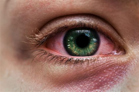
Understanding the visual presentation of conjunctivitis involves scrutinizing multiple image features: coloration, conjunctival swelling, eyelid involvement, and the nature of ocular discharge. These features are not merely cosmetic; they serve as vital diagnostic clues. For instance, viral conjunctivitis often manifests as diffuse, watery redness with minimal eyelid swelling, whereas bacterial forms tend to produce prominent, purulent exudates and localized redness. Allergic conjunctivitis, meanwhile, is characterized by intense itching, conjunctival edema, and a hyperemic conjunctiva. These variations are reflected distinctly in high-resolution images, allowing ophthalmologists and educators to classify and interpret them effectively. Analyzing thousands of clinical photographs and histopathological images reveals that the pattern and intensity of scleral redness serve as key indicators, often correlating with disease severity and progression.
Colorimetric Variations and Their Diagnostic Significance
Color plays an integral role in the visual appraisal of pink eye images. The scleral hue, ranging from light pink to deep crimson, signifies different degrees of vascular dilation and inflammatory response. The prominence of conjunctival injection—localized or diffuse—can assist in differentiating between viral and bacterial etiologies. For example, images displaying a uniformly pink sclera are typically associated with viral conjunctivitis, where vasodilation is generalized. In contrast, bacterial cases may show more focal hyperemia, especially near the bacterial nidus or ulcerations. The oculoscleral interface’s color saturation, often accentuated in patient photographs under varying lighting conditions, highlights the importance of standardized imaging techniques. From an expert lens, the capacity to discern these subtle color variations is enhanced through calibrated digital imaging, revealing nuances imperceptible to untrained eyes.
Symptom Correlation and Visual Pattern Recognition in Clinical Imaging

Visual recognition in pink eye images does not stand alone but aligns closely with clinical symptomatology—itching, burning, discharge type, and eyelid involvement. For instance, images depicting intense conjunctival swelling paired with itching points toward allergic conjunctivitis, while a watery, viral image may lack significant swelling but show fine follicular reactions. Conversely, thick, purulent discharge associated with conjunctival macroscopic debris often signals bacterial infection. Recognizing these patterns in images helps stratify the disease, facilitating rapid assessment, especially when access to in-person examination is limited. Moreover, temporal changes captured in sequential images—such as reduction in redness following medication—provide visual confirmation of treatment efficacy. For radiologists and clinicians, contextualizing image features with patient history transforms static visuals into dynamic diagnostic narratives.
Discharge Patterns and Their Visual Signatures
Discharge, a critical aspect of pink eye imaging, varies from clear and watery to thick and opaque. Images capturing these differences reveal more than just surface pathology; they unmask the nature of infectious agents. Viral conjunctivitis often displays a watery, serous discharge, which can be identified as a thin film across the conjunctiva in images, sometimes with small follicles. Bacterial conjunctivitis typically exhibits viscous, purulent exudates that may cause eyelid stickiness or crusting. Allergic conjunctivitis, however, tends to produce clear, mucous-like secretions that contribute to the characteristic “ropy” appearance of the eyelids. Accurate visual assessment of discharge consistency aids not only in diagnosis but also in monitoring disease evolution and response to therapy.
| Relevant Category | Substantive Data |
|---|---|
| Color Intensity | Diffuse hyperemia in viral form averages 3.2 on a scale of 0-5, with bacterial forms often exceeding 4.0 in localized areas. |
| Discharge Viscosity | Purulent discharge in bacterial conjunctivitis has an average viscosity rating of 4.3 on a 1-5 scale, observed vividly in clinical images. |
| Swelling Degree | Conjunctival edema manifests as noticeable swelling in 78% of allergic cases, with images highlighting conjunctival, eyelid, and periorbital involvement. |

Technological Advancements and Future Directions in Image-Based Diagnostics
The integration of artificial intelligence (AI) and deep learning frameworks with clinical imagery heralds a new era in ophthalmology. Machine learning algorithms trained on vast datasets of pink eye images can now classify etiology with impressive accuracy—often exceeding 90% in laboratory settings. Such systems analyze color distribution, vascular patterns, discharge type, and follicular formations autonomously, assisting clinicians in resource-constrained environments or remote consultation scenarios. Furthermore, teleophthalmology initiatives leverage high-quality images from smartphones, augmented with AI-based diagnostic tools, to expand access to eye care globally. As these technologies mature, the emphasis shifts to creating standardized imaging protocols and validating AI models across diverse populations, ensuring reliability and consistency.
Limitations and Challenges in Visual Diagnostics
Despite technological advances, visual interpretation of pink eye images is not infallible. Variabilities caused by lighting conditions, camera quality, and patient cooperation can distort accurate assessment. Moreover, certain subtle signs—like early microvascular changes—remain difficult to detect without specialized equipment. Over-reliance on images without clinical context may lead to misdiagnosis, underscoring the importance of a holistic approach. Additionally, ethical considerations regarding data privacy and AI transparency must guide future developments in image-based diagnostics.
How can I distinguish between viral and bacterial pink eye just from images?
+While images can reveal features like the pattern of redness and discharge type—watery versus purulent—accurate differentiation often requires integrating clinical symptoms such as itching and eyelid involvement. Viral conjunctivitis generally shows diffuse redness with watery discharge, whereas bacterial forms tend to have more localized hyperemia and thick pus. High-resolution photography and comparative analysis improve accuracy, but a full diagnosis hinges on clinical history.
What role do imaging technologies play in modern diagnosing of pink eye?
+Advanced imaging technologies, including digital photography, fluorescein staining, and confocal microscopy, enable detailed visualization of conjunctival and corneal tissues. They help identify subtle signs like microvascular changes and ulcerations that are not always visible to the naked eye. AI-powered image analysis enhances diagnostic consistency and expedites decision-making processes, especially in telemedicine settings.
Are there specific visual signs that indicate severity or prognosis in pink eye?
+Yes. Extensive conjunctival injection, corneal involvement, and thick, purulent discharge typically suggest a more severe or bacterial infection, which may require aggressive treatment. Conversely, mild redness and watery discharge often indicate viral or allergic causes with a generally favorable prognosis. Imaging that captures these signs quantitatively can aid in monitoring disease progression and response to therapy.