The human brain, often described as the most intricate organ in the body, holds mysteries that continue to fascinate neuroscientists and neuroanatomists worldwide. Among its many features, the longitudinal fissure stands out as a prominent and defining anatomical landmark, separating the brain's two hemispheres—the left and the right. Despite its apparent simplicity, this fissure plays a crucial role in our understanding of brain organization, neural communication, and functional specialization. Over the decades, the exploration of the longitudinal fissure has helped shape our comprehension of hemispheric lateralization, interhemispheric connectivity, and the structural differences that underlie various cognitive processes. This comprehensive guide aims to dissect the anatomy, function, clinical significance, and ongoing debates surrounding the longitudinal fissure, presenting balanced perspectives while synthesizing current scientific consensus.
Understanding the Anatomy and Development of the Longitudinal Fissure

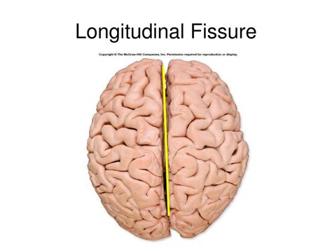
The longitudinal fissure, also known as the cerebral fissure, is a deep groove that runs along the median plane of the brain, effectively dividing the cerebrum into two nearly symmetrical halves. Its formation originates during the early stages of embryonic neural development, specifically around the 8th to 10th week of gestation, as the telencephalon undergoes lateral expansion and cortical proliferation.
Structurally, the fissure extends from the inferior margins of the brain, beginning at the frontal pole and continuing posteriorly towards the occipital lobe, terminating near the cerebellum. It encompasses not merely a superficial groove but also underlies critical structures such as the falx cerebri—a dural septum that provides stability and houses critical venous sinuses. This dural partition ensures that the hemispheres are physically separated, reducing mechanical interference during movement and providing space for vascular and neural organization.
Macroscopically, the fissure is characterized by its depth and the presence of the cingulate gyrus, which lies superiorly, and the corpus callosum—the largest white matter tract connecting both hemispheres—residing deep within the fissure. The development of this structure is closely linked with the overall lateralization of brain functions and the interhemispheric integration of neural signals.
| Relevant Category | Substantive Data |
|---|---|
| Depth of fissure | Approximately 4-6 mm on average, with variability based on individual differences |
| Extension length | Approximately 13-15 cm from frontal pole to occipital pole in adults |
| Primary associated structures | Falx cerebri, corpus callosum, cingulate gyrus |

The Functional Significance and Hemispheric Specialization

While at first glance the longitudinal fissure appears to be a mere physical divider, it encapsulates the complexity of how the brain segregates and integrates functions across hemispheres. The division allows each hemisphere to develop specialized neural circuits, leading to hemispheric dominance in tasks like language, spatial reasoning, and emotional processing.
Hemispheric Lateralization and the Role of the Fissure
The concept of hemispheric lateralization describes the tendency for certain cognitive functions to be predominantly managed by one hemisphere. For example, language centers, such as Broca’s and Wernicke’s areas, are typically localized in the left hemisphere in right-handed individuals. Conversely, the right hemisphere often processes visuospatial information and aspects of emotion recognition.
The physical separation enforced by the longitudinal fissure facilitates this functional segregation. It enables each hemisphere to operate semi-independently during complex tasks, minimizing cross-hemispheric interference—though the corpus callosum regulates interhemispheric communication essential for integrated processing.
Neuroimaging studies, including functional MRI (fMRI), have demonstrated that tasks activate distinct but interconnected regions across the two hemispheres. This interplay relies heavily on the structural integrity of the fissure and associated commissural fibers.
| Related Metric | Data and Context |
|---|---|
| Degree of lateralization | Approximately 95% of right-handed individuals exhibit left lateralization for language tasks |
| Corpus callosum connectivity | Contains roughly 200 million axons facilitating interhemispheric transfer |
Clinical and Surgical Implications of the Longitudinal Fissure
The prominence of the longitudinal fissure extends beyond anatomy; it bears significance in clinical neurology, neurosurgery, and neuropsychology. Surgical approaches, particularly those involving corpus callosotomy for severe epilepsy, exploit the fissure as a natural boundary to access and modify neural circuits while minimizing damage to surrounding tissues.
Surgical Access and Risks
Procedures such as corpus callosotomy involve sectioning the corpus callosum—primarily within the longitudinal fissure—to prevent epileptic discharges from propagating across hemispheres. The anatomical clarity provided by the fissure facilitates this procedure, though it necessitates precise navigation to avoid critical structures like the falx cerebri and sagittal sinus.
Risks include disconnection syndromes, anosognosia, and disruptions in interhemispheric transfer functions. Postoperative management must consider potential behavioral and cognitive changes, emphasizing the importance of detailed preoperative mapping.
| Surgical Metric | Data and Context |
|---|---|
| Success rate | Approximately 60-70% in reducing severe seizure frequency |
| Common complications | Disconnection syndromes (~10%), transient deficits, neurological deficits |
Contemporary Debates and Future Directions
Despite long-standing knowledge, debates persist regarding the exact functional implications of fissure morphology and its variability among individuals. Some scholars argue that the fissure’s size and shape may correlate with neurodevelopmental disorders such as autism spectrum disorder (ASD) or schizophrenia, although conclusive evidence remains elusive.
The Variability and Plasticity of the Fissure
Recent neuroimaging research suggests that the morphology of the longitudinal fissure may exhibit considerable individual differences, influenced by genetic and environmental factors. Such variability could influence interhemispheric communication efficiency or predispose individuals to certain neuropsychological conditions.
Moreover, neural plasticity—the brain’s ability to compensate for structural differences or injuries—raises questions about whether the fissure’s characteristics can adapt or influence functional reorganization over time.
| Aspect | Current Understanding |
|---|---|
| Variability in fissure size | Ranges widely; some individuals exhibit deeper or shallower fissures |
| Link to neurodevelopmental disorders | Suggestive but not conclusive evidence |
| Potential for plasticity | Limited evidence; more research needed |
Synthesis and Concluding Perspectives

The longitudinal fissure exemplifies how a seemingly simple anatomical feature is imbued with profound functional and clinical significance. It constitutes a critical boundary that supports the lateralization of brain functions, facilitates targeted neurosurgical interventions, and reflects the developmental intricacies encoded within our neural architecture. While debates concerning its variability and functional implications continue, integration of neuroimaging, neurogenetics, and clinical data enriches our understanding, promising more tailored and effective interventions in neurology and psychiatric care.
Ultimately, appreciating the complexity of the longitudinal fissure enriches our broader understanding of neural organization, bridging the gap between structure and function. As neuroscience advances, this fissure will remain a focal point for unraveling the mysteries of hemispheric specialization and interhemispheric communication.
What is the primary function of the longitudinal fissure?
+The longitudinal fissure primarily separates the two cerebral hemispheres, allowing for structural and functional differentiation, while facilitating interhemispheric communication through the corpus callosum.
How does the variability in the fissure’s anatomy affect neurological health?
+Variations in fissure size or morphology may influence interhemispheric connectivity and potentially predispose individuals to neurodevelopmental or neuropsychiatric conditions, though more research is needed to establish definitive links.
Can surgical intervention involving the fissure be performed safely?
+Yes, procedures like corpus callosotomy leverage the fissure’s anatomy for targeted intervention. Success depends on precise imaging, surgical expertise, and careful postoperative management to mitigate disconnection syndromes.